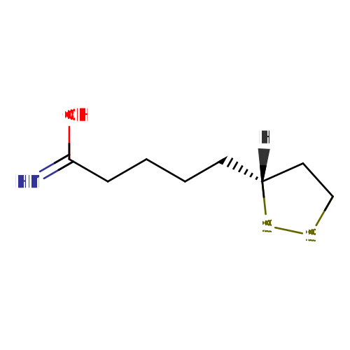
Lysozyme (CED0000854)

Record Information
Version1.0
Creation Date2016-05-25 20:52:20 UTC
Update Date2026-04-14 19:21:16 UTC
Accession NumberCHEM023157
Identification
Common NameLysozyme
ClassSmall Molecule
Description
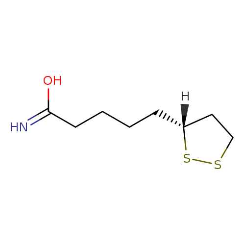
Lysozyme is an organoheterocyclic compound with the chemical formula C8H15NOS2 and an average molecular weight of 205.33 g/mol. Lysozyme belongs to the lipoamides, a subclass of dithiolanes within the organic compounds. In industrial applications, this compound is utilized as a conditioner and softener. It has been recorded as being found in or released from amplifiers within electrical and electronic equipment.
Contaminant TypeNot Available
Chemical Structure
Synonyms
ValueSource
5-(1,2-Dithiolan-3-yl)-pentanamideMetaCyc
DL-LipoamideMetaCyc
6,8-Dithiooctanoic amideMetaCyc
Chemical FormulaC8H15NOS2
Average Molecular Mass205.330 g/mol
Monoisotopic Mass205.060 g/mol
CAS Registry Number9001-63-2
IUPAC Name5-[(3R)-1,2-dithiolan-3-yl]pentanimidic acid
Traditional Namelipoamide
SMILES[H][C@@]1(CCCCC(O)=N)CCSS1
InChI IdentifierInChI=1S/C8H15NOS2/c9-8(10)4-2-1-3-7-5-6-11-12-7/h7H,1-6H2,(H2,9,10)/t7-/m1/s1
InChI KeyFCCDDURTIIUXBY-SSDOTTSWSA-N
Chemical Taxonomy
Description Belongs to the class of organic compounds known as lipoamides. Lipoamides are compounds containing a lipoamide moiety, which consists of a pentanamide attached to the C3 carbon atom of a 1,2-dithiolane ring.
KingdomOrganic compounds
Super ClassOrganoheterocyclic compounds
ClassDithiolanes
Sub ClassLipoamides
Direct ParentLipoamides
Alternative Parents
Substituents
  • Lipoamide
  • Fatty amide
  • Fatty acyl
  • 1,2-dithiolane
  • Carboxamide group
  • Organic disulfide
  • Primary carboxylic acid amide
  • Carboxylic acid derivative
  • Carbonyl group
  • Organooxygen compound
  • Organonitrogen compound
  • Hydrocarbon derivative
  • Organic oxide
  • Organopnictogen compound
  • Organic oxygen compound
  • Organic nitrogen compound
  • Aliphatic heteromonocyclic compound
Molecular FrameworkAliphatic heteromonocyclic compounds
External Descriptors
Biological Properties
StatusDetected and Not Quantified
OriginNot Available
Cellular LocationsNot Available
Biofluid LocationsNot Available
Tissue LocationsNot Available
ApplicationsNot Available
Biological RolesNot Available
Chemical RolesNot Available
Organoleptic EffectsNot Available
Physical Properties
StateNot Available
AppearanceNot Available
Experimental Properties
PropertyValue
Melting PointNot Available
Boiling PointNot Available
SolubilityNot Available
Predicted Properties
PropertyValueSource
Water Solubility0.11 g/LALOGPS
logP2.47ALOGPS
logP-0.52ChemAxon
logS-3.3ALOGPS
pKa (Strongest Acidic)-0.064ChemAxon
pKa (Strongest Basic)13.21ChemAxon
Physiological Charge0ChemAxon
Hydrogen Acceptor Count2ChemAxon
Hydrogen Donor Count2ChemAxon
Polar Surface Area44.08 ŲChemAxon
Rotatable Bond Count5ChemAxon
Refractivity67.13 m³·mol⁻¹ChemAxon
Polarizability22.05 ųChemAxon
Number of Rings1ChemAxon
BioavailabilityYesChemAxon
Rule of FiveYesChemAxon
Ghose FilterNoChemAxon
Veber's RuleNoChemAxon
MDDR-like RuleNoChemAxon
Spectra
Spectra
Spectrum TypeDescriptionSplash KeyDeposition DateView
Predicted LC-MS/MSPredicted LC-MS/MS Spectrumsplash10-0a4r-0940000000-d79e1aa52057e567d71fNot AvailableView Spectrum
Predicted LC-MS/MSPredicted LC-MS/MS Spectrumsplash10-052r-3900000000-abd8148048c5f282d889Not AvailableView Spectrum
Predicted LC-MS/MSPredicted LC-MS/MS Spectrumsplash10-0adr-9500000000-9769c619660ab6af46d2Not AvailableView Spectrum
Predicted LC-MS/MSPredicted LC-MS/MS Spectrumsplash10-0zml-1920000000-79d57d488cb8fbc804a3Not AvailableView Spectrum
Predicted LC-MS/MSPredicted LC-MS/MS Spectrumsplash10-0fdo-4910000000-d264eaf6e242c36bc563Not AvailableView Spectrum
Predicted LC-MS/MSPredicted LC-MS/MS Spectrumsplash10-0006-9000000000-289d2f20046f7819a7caNot AvailableView Spectrum
Predicted LC-MS/MSPredicted LC-MS/MS Spectrumsplash10-0a4i-0690000000-db32b4efef6d4d06e799Not AvailableView Spectrum
Predicted LC-MS/MSPredicted LC-MS/MS Spectrumsplash10-03di-1900000000-c09c2f759aca294a35f0Not AvailableView Spectrum
Predicted LC-MS/MSPredicted LC-MS/MS Spectrumsplash10-0ika-5900000000-b388d7bb80b06afcd3b9Not AvailableView Spectrum
Predicted LC-MS/MSPredicted LC-MS/MS Spectrumsplash10-0udi-0090000000-026e73840b34e98efd4bNot AvailableView Spectrum
Predicted LC-MS/MSPredicted LC-MS/MS Spectrumsplash10-0zfr-9770000000-2f59ecc3f1f23cd58faaNot AvailableView Spectrum
Predicted LC-MS/MSPredicted LC-MS/MS Spectrumsplash10-0006-9000000000-1e1344f874294cd76bceNot AvailableView Spectrum
Toxicity Profile
Mechanism of ToxicityNot Available
Carcinogenicity (IARC Classification)Not Available
Minimum Risk LevelNot Available
SymptomsNot Available
TreatmentNot Available
Toxicity Values
Toxicity ValueUnitValue RangeOrganismDose DescriptorRoute of ExposurePredicted or ExperimentalReference
722.0Log mg/kg[410:1300]RatLD50oralpredictedNot Available
Health Effects
Health EffectRelationshipDirectionReference
Exposure Sources
Source IDSourceSectorReference
505AmplifiersElectrical & Electronic EquipmentNot Available
Pathways
0 pathways

No pathways found

No metabolic pathways have been associated with this synthetic chemical

Targets
StructureProteinUniProt IDOrganismRelationshipDetails
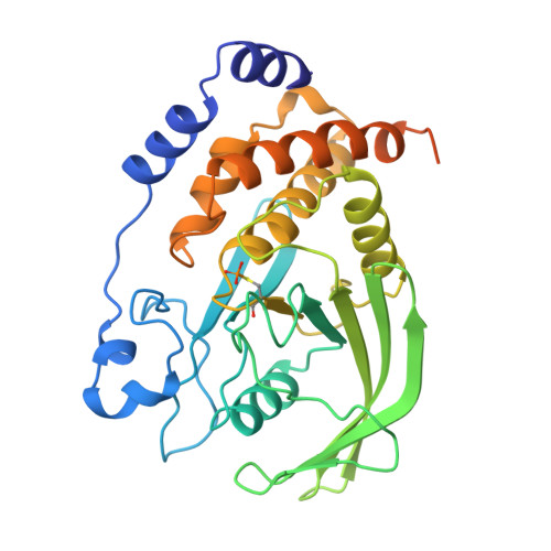
Acetylcholinesterase structureClick to view 3D structureAcetylcholinesteraseP22303HumansPredicted (SEA)1969.9
Prostaglandin G/H synthase 2 structureClick to view 3D structureProstaglandin G/H synthase 2P35354HumansPredicted (SEA)1395.21
Nuclear receptor subfamily 1 group I member 2 structureClick to view 3D structureNuclear receptor subfamily 1 group I member 2O75469HumansPredicted (SEA)614.23
Click to view 3D structureAlpha-1A adrenergic receptorP43140Rattus norvegicusPredicted (SEA)1460.3
Click to view 3D structureAlpha-1B adrenergic receptorP15823Rattus norvegicusPredicted (SEA)900.404
Click to view 3D structureProtein TatP04608Human immunodeficiency virus type 1 group M subtype B (isolate HXB2)(HIV-1)Predicted (SEA)554.442
Click to view 3D structureAlpha-1D adrenergic receptorP23944Rattus norvegicusPredicted (SEA)1247.77
Tyrosine-protein phosphatase non-receptor type 1 structureClick to view 3D structureTyrosine-protein phosphatase non-receptor type 1P18031HumansPredicted (SEA)2105.12
Click to view 3D structureBifunctional protein GlmUP9WMN3Mycobacterium tuberculosisPredicted (SEA)0.0778492
Fibronectin structureClick to view 3D structureFibronectinP02751HumansPredicted (SEA)4.7488
Prostaglandin E2 receptor EP2 subtype structureClick to view 3D structureProstaglandin E2 receptor EP2 subtypeP43116HumansPredicted (SEA)3983.16
Click to view 3D structureProstaglandin E2 receptor EP1 subtypeP34995HumansPredicted (SEA)4275.4
Thromboxane A2 receptor structureClick to view 3D structureThromboxane A2 receptorP21731HumansPredicted (SEA)5321.69
D(2) dopamine receptor structureClick to view 3D structureD(2) dopamine receptorP14416HumansPredicted (SEA)7568.35
5-hydroxytryptamine receptor 1A structureClick to view 3D structure5-hydroxytryptamine receptor 1AP08908HumansPredicted (SEA)7544.76
Prostacyclin receptor structureClick to view 3D structureProstacyclin receptorP43119HumansPredicted (SEA)5158.13
Prostaglandin E2 receptor EP3 subtype structureClick to view 3D structureProstaglandin E2 receptor EP3 subtypeP43115HumansPredicted (SEA)5923.7
Tyrosine-protein phosphatase non-receptor type 2 structureClick to view 3D structureTyrosine-protein phosphatase non-receptor type 2P17706HumansPredicted (SEA)4075.95
Peroxisome proliferator-activated receptor alpha structureClick to view 3D structurePeroxisome proliferator-activated receptor alphaQ07869HumansPredicted (SEA)6522.29
Peroxisome proliferator-activated receptor delta structureClick to view 3D structurePeroxisome proliferator-activated receptor deltaQ03181HumansPredicted (SEA)6345.18
Peroxisome proliferator-activated receptor gamma structureClick to view 3D structurePeroxisome proliferator-activated receptor gammaP37231HumansPredicted (SEA)7135.04
Click to view 3D structureAlpha-1D adrenergic receptorP25100HumansPredicted (SEA)7427.21
Mitofusin-2 structureClick to view 3D structureMitofusin-2O95140HumansPredicted (SEA)1389.14
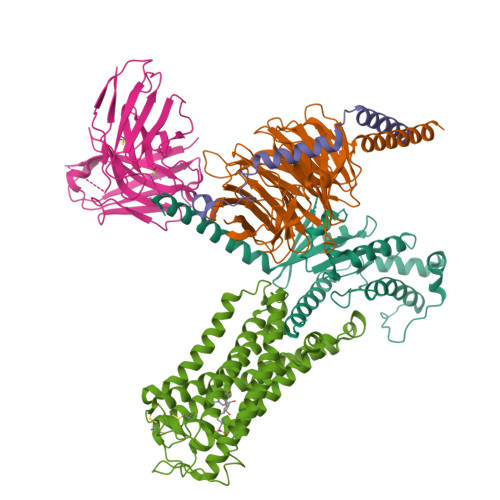
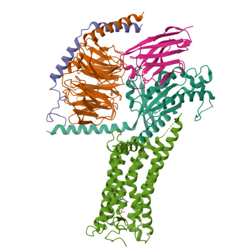
Guanine nucleotide-binding protein G(i) subunit alpha-1 structureClick to view 3D structureGuanine nucleotide-binding protein G(i) subunit alpha-1P63096HumansPredicted (SEA)347.908
Prostaglandin E2 receptor EP4 subtype structureClick to view 3D structureProstaglandin E2 receptor EP4 subtypeP35408HumansPredicted (SEA)6710.37
Concentrations
Not Available
External Links
DrugBank IDNot Available
HMDB IDHMDB0304823
FooDB IDFDB000540
Phenol Explorer IDNot Available
KNApSAcK IDNot Available
BiGG IDNot Available
BioCyc IDLIPOAMIDE
METLIN IDNot Available
PDB IDNot Available
Wikipedia LinkNot Available
Chemspider ID5360246
ChEBI ID83094
PubChem Compound IDNot Available
Kegg Compound IDNot Available
YMDB IDNot Available
ECMDB IDNot Available
References
Synthesis ReferenceNot Available
MSDSNot Available
General References