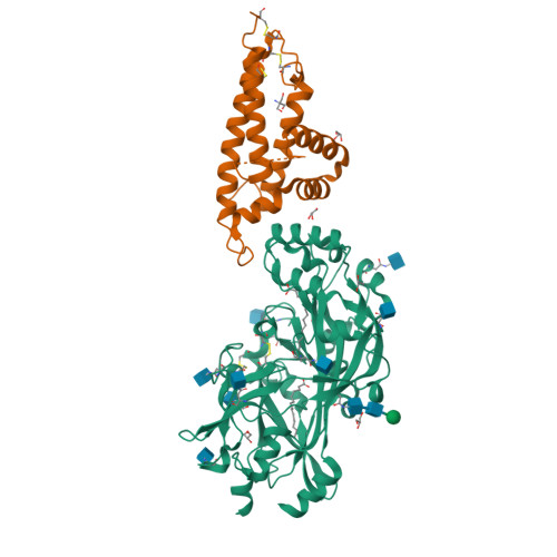
 Click to view 3D structure Click to view 3D structure | Platelet glycoprotein 4 | P16671 | Humans | Predicted (SEA) | 5.3716 |
 Click to view 3D structure Click to view 3D structure | 1-phosphatidylinositol 4,5-bisphosphate phosphodiesterase delta-1 | P10688 | Rattus norvegicus | Predicted (SEA) | 2.56902 |
| Click to view 3D structure | 1-phosphatidylinositol 4,5-bisphosphate phosphodiesterase delta-1 | P51178 | Humans | Predicted (SEA) | 2.56902 |
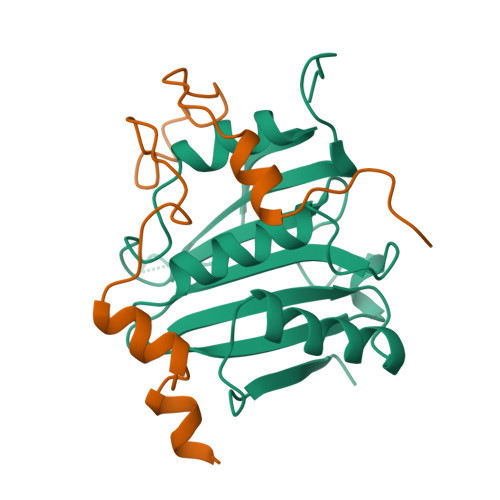 Click to view 3D structure Click to view 3D structure | GRB10-interacting GYF protein 2 | Q6Y7W6 | Humans | Predicted (SEA) | 10.3539 |
| Click to view 3D structure | Biotin--protein ligase | P50747 | Humans | Predicted (SEA) | 2.95827 |
| Click to view 3D structure | BirA bifunctional protein | P96884 | Mycobacterium tuberculosis (strain CDC 1551 / Oshkosh) | Predicted (SEA) | 14.9471 |
| Click to view 3D structure | Biotin--[acetyl-CoA-carboxylase] ligase | I6YFP0 | Mycobacterium tuberculosis (strain ATCC 25618 / H37Rv) | Predicted (SEA) | 27.87 |
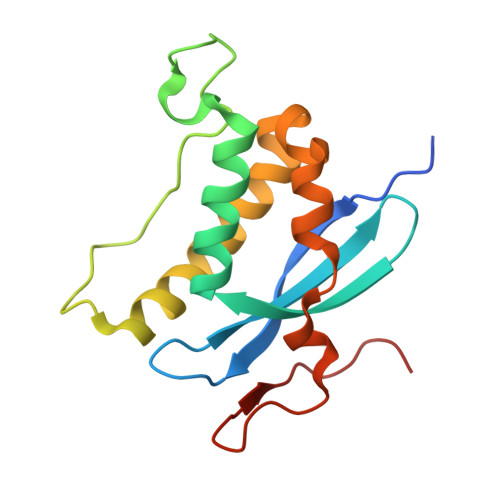 Click to view 3D structure Click to view 3D structure | NADPH oxidase organizer 1 | Q8NFA2 | Humans | Predicted (SEA) | 8.17417 |
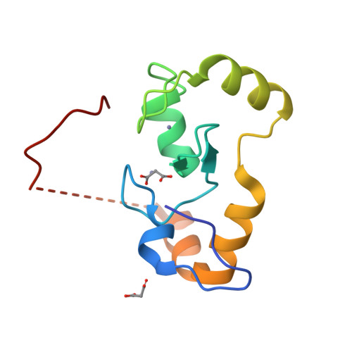 Click to view 3D structure Click to view 3D structure | E3 ubiquitin-protein ligase RNF168 | Q8IYW5 | Humans | Predicted (SEA) | 18.3724 |
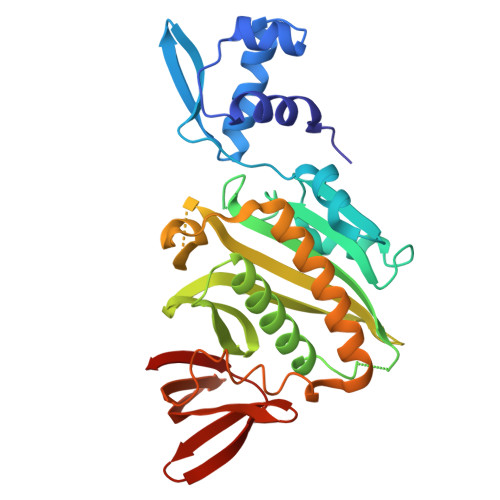 Click to view 3D structure Click to view 3D structure | Bifunctional ligase/repressor BirA | P06709 | Escherichia coli (strain K12) | Predicted (SEA) | 44.2962 |
| Click to view 3D structure | Nonstructural protein 1 | Q194T2 | Influenza A virus | Predicted (SEA) | 453.394 |
| Click to view 3D structure | Bifunctional ligase/repressor BirA | A0A0E7NXW5 | Staphylococcus aureus | Predicted (SEA) | 10.5875 |
 Click to view 3D structure Click to view 3D structure | Streptavidin | P22629 | Streptomyces avidinii | Predicted (SEA) | 3.19182 |
| Click to view 3D structure | Lymphocyte antigen 6K | Q17RY6 | Humans | Predicted (SEA) | 57.9198 |
| Click to view 3D structure | Cytohesin-3 | P97696 | Rattus norvegicus | Predicted (SEA) | 3.11397 |
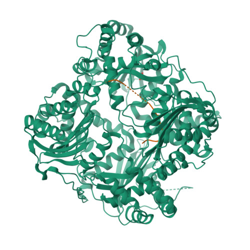 Click to view 3D structure Click to view 3D structure | Insulin-degrading enzyme | P14735 | Humans | Predicted (SEA) | 273.874 |
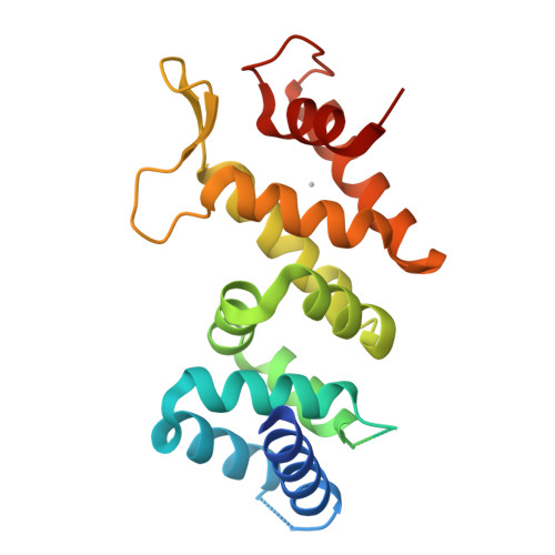 Click to view 3D structure Click to view 3D structure | Fanconi anemia group F protein | Q9NPI8 | Humans | Predicted (SEA) | 5.91654 |
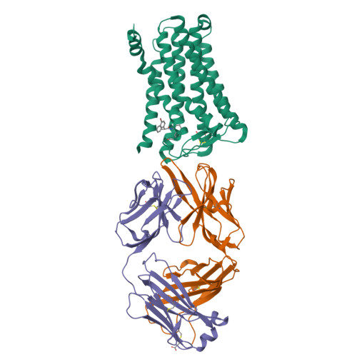 Click to view 3D structure Click to view 3D structure | Prostaglandin E2 receptor EP4 subtype | P35408 | Humans | Predicted (SEA) | 3602.47 |
 Click to view 3D structure Click to view 3D structure | Prostaglandin E2 receptor EP2 subtype | P43116 | Humans | Predicted (SEA) | 3586.98 |
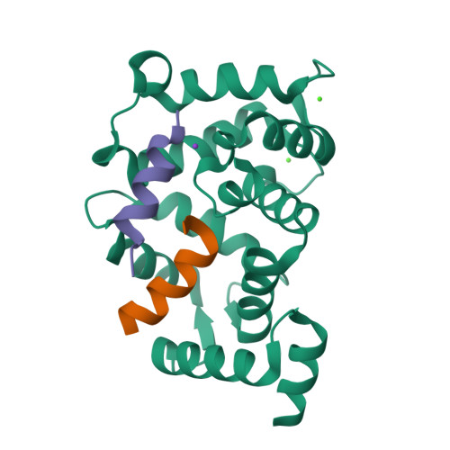 Click to view 3D structure Click to view 3D structure | D(2) dopamine receptor | P14416 | Humans | Predicted (SEA) | 7087.32 |
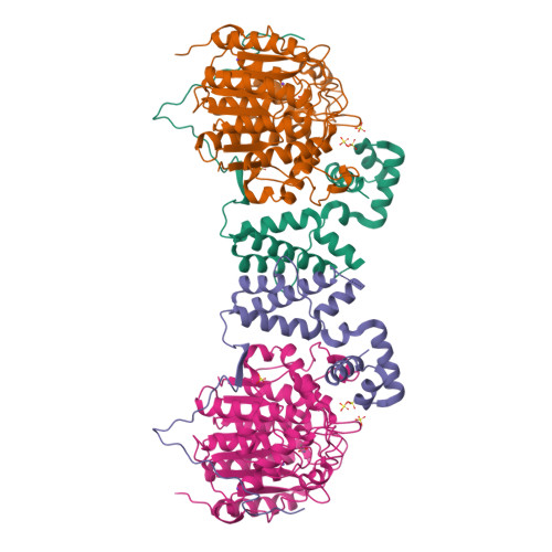 Click to view 3D structure Click to view 3D structure | Histone deacetylase 1 | Q13547 | Humans | Predicted (SEA) | 6590.25 |
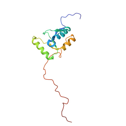 Click to view 3D structure Click to view 3D structure | DNA-directed DNA/RNA polymerase mu | Q9NP87 | Humans | Predicted (SEA) | 204.51 |
| Click to view 3D structure | DNA nucleotidylexotransferase | P06526 | Bos taurus | Predicted (SEA) | 204.51 |
| Click to view 3D structure | Prostaglandin E2 receptor EP1 subtype | P34995 | Humans | Predicted (SEA) | 3754.2 |
| Click to view 3D structure | Cytochrome P450 2C23 | P24470 | Rattus norvegicus | Predicted (SEA) | 31.1397 |